A CHALLENGING CASE OF RECURRING MULTIPLE INTRAOSSEOUS BONE HEMANGIOMA IN YOUNG FEMALE WITH A LONG TERM 10 YEAR FOLLOW-UP
AKSHAY P¹, JOHN T JOHN2, JOICE VARGHESE M J3, RENJIT JOHN MATHEW4, SHINAS B SALAM5, DAVID JOSEPH6
Abstract
Background:
Intraosseous hemangioma is a rare benign vascular tumor, accounting for less than 1% of all bone tumors. Although it commonly affects the vertebrae and skull, long-bone or rib involvement is distinctly uncommon, and multifocal recurrence is exceedingly rare.
Case Presentation:
We report the case of a young female with recurrent multifocal intraosseous hemangiomas initially involving the right 9th rib, followed years later by progressive lesions of the right proximal femur, glenoid bone, and iliac bone. The patient underwent interventions including bone excision, biopsy, dynamic hip screw fixation, and iliac–fibular strut bone grafting. Despite benign histopathology, the lesions demonstrated aggressive radiologic progression over a decade of follow-up.
Conclusion:
This case highlights the diagnostic and therapeutic challenges of multifocal recurrent intraosseous hemangiomas. Long-term surveillance using multimodal imaging is essential for timely detection of recurrence or dissemination, and a multidisciplinary approach is imperative in managing structurally threatening lesions.
Introduction
Intraosseous hemangioma is an uncommon benign vascular tumor arising from blood vessels within the bone. It represents less than 1% of all osseous neoplasms. While vertebrae and craniofacial bones are the most frequently affected sites, involvement of the appendicular skeleton is rare. The natural history is variable, ranging from incidental findings to symptomatic, expansile, or destructive lesions. Multifocal lesions or disseminated involvement remain exceedingly unusual and pose significant diagnostic and therapeutic dilemmas.
We present a rare case of a young female with recurrent, multifocal intraosseous hemangiomas affecting multiple skeletal sites over a span of ten years, emphasizing the importance of vigilant long-term follow-up.
Case Presentation
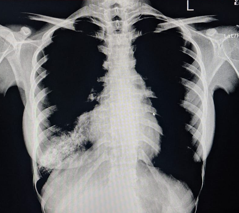
A young 23 year old female first presented in 2015 with complaints of right-sided chest pain and swelling. A thorough clinico-radiological evaluation was done and general surgeon consultation sought. Imaging revealed an expansile lytic lesion of the right 9th rib (Fig 1). Other long bone x-ray evaluation was done and found no abnormalities. We planned for excision biopsy of 9th rib and patient underwent surgical excision (Fig 2). Histopathological examination confirmed a benign intraosseous hemangioma(Fig 3). Post-operative recovery was uneventful. 6 monthly followed by yearly radiological follow-up was being done till pre-covid period. Patient lost to follow-up during 2020-2022 Covid-19 period.
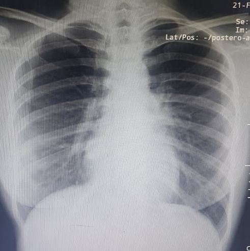
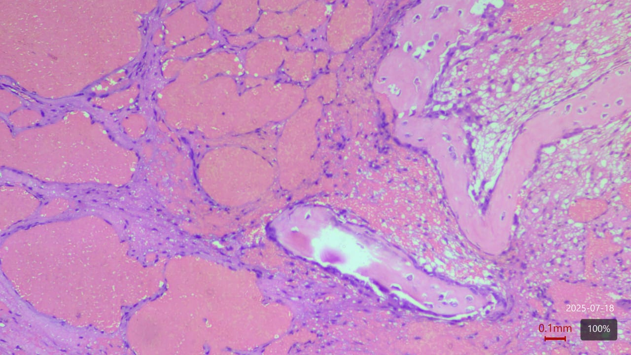
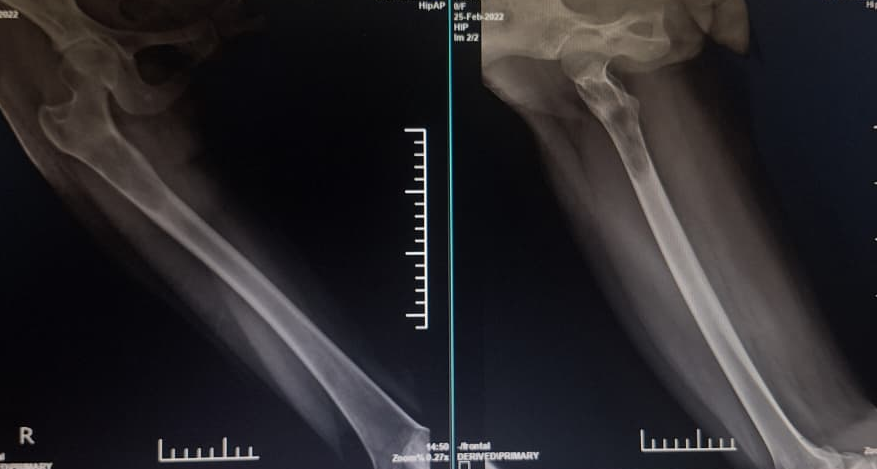
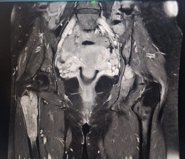
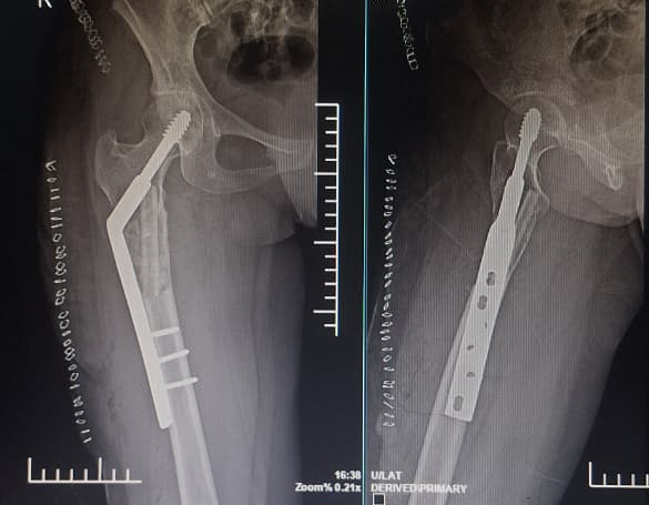
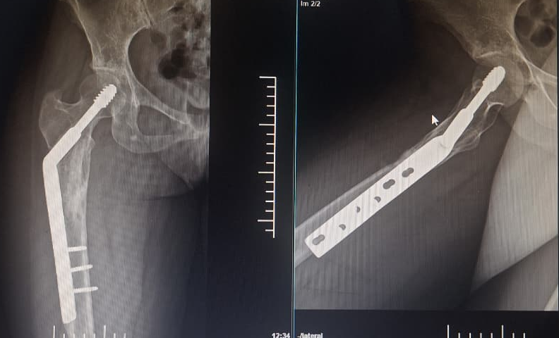
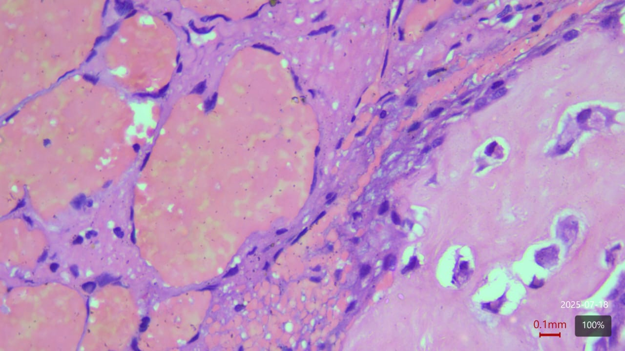
In 2022, the patient presented with progressive pain and restricted range of motion of the right hip following minor trauma. She was re-evaluated clinically and imaging was done. Radiographs (Fig 4 ) and MRI (Fig 5) revealed an expansile lytic lesion of the right proximal femur consistent with intraosseous hemangioma. Due to risk of pathological fracture we planned for fixation of right proximal femur. She underwent right proximal femoral bone excision biopsy and dynamic hip screw fixation with iliac–fibular strut bone grafting (Fig 6). Post-operative period was uneventful. Started partial weight bearing after 45 days followed by full weight bearing by 90 days. Very good radiological incorporation of graft noted and fixation was stable radiologically (Fig 7). Histopathology report suggestive of benign bone hemangiomatous tissue without any features of malignancy (Fig 8).
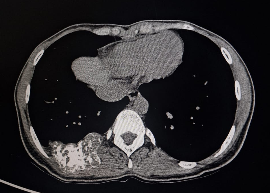
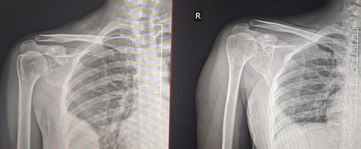
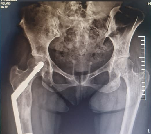
Subsequent follow-up imaging from 2023 to 2025 showed continued multifocal progression. A rapidly enlarging lytic lesion was identified in the right glenoid bone found to regress in follow-up x-ray (Fig 9). An additional lytic foci appeared in the right iliac bone in latest follow-up of 2025 ( Fig 10) when she presented with complaints of mild pain from right groin. Despite the aggressive radiologic appearance, repeat biopsy demonstrated benign hemangiomatous tissue without features of malignancy, hence we did not plan for any intervention.
The patient is kept in long-term surveillance, with stable postoperative hip fixation and no current functional impairment.
Investigations
Radiographic Findings
- 2015: Expansile lytic lesion of right 9th rib with cortical thinning (Fig 1).
- 2022: Lytic right proximal femur lesion with risk of pathological fracture (Fig 4).
- 2023–2025:
- Rapidly progressive right glenoid bone lesion regressing in follow-up (Fig 9).
- Lytic foci in the right iliac bone (Fig 10).
- Postoperative consolidation of proximal femoral graft (Fig 7).
MRI
MRI of the proximal femur showed characteristic high-flow vascular channels consistent with intraosseous hemangioma (Fig 5).
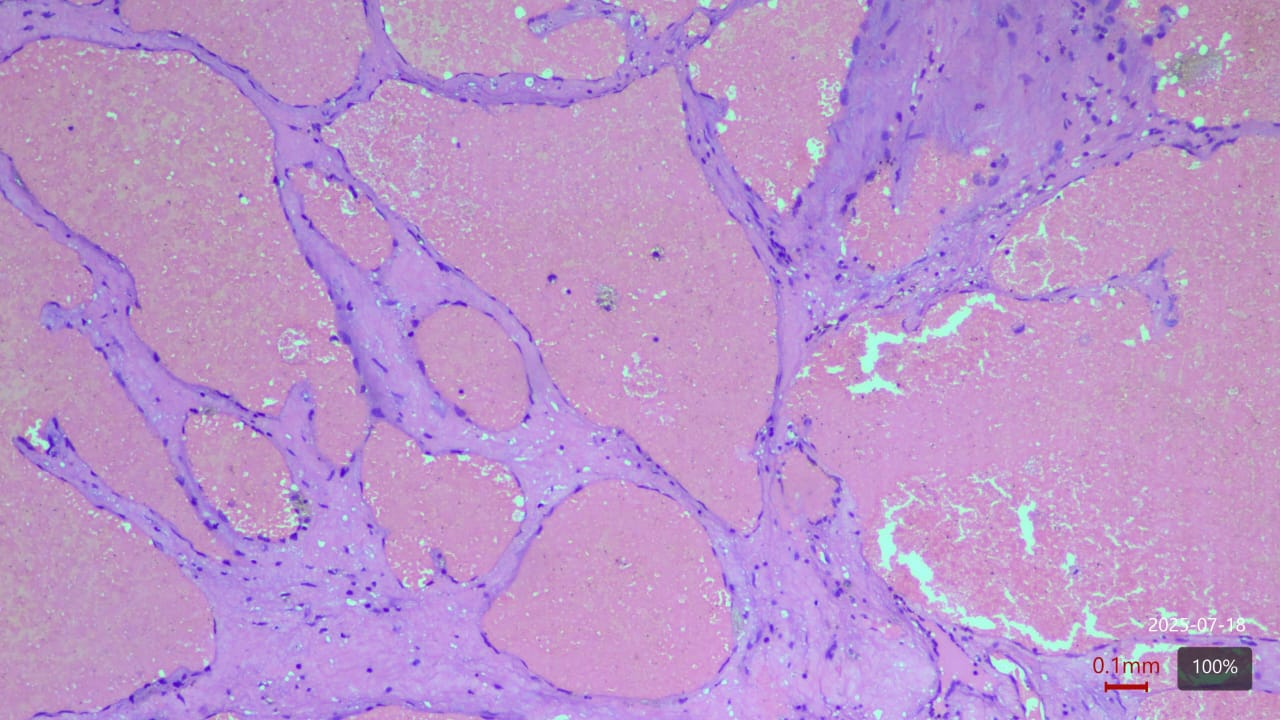
Histopathology
Biopsies demonstrated benign vascular channels lined by flattened endothelial cells with no atypia or malignant transformation. Despite the benign appearance, radiological progression persisted (Fig 8).
Intervention
The patient underwent right proximal femur bone excision biopsy, dynamic hip screw fixation, and iliac–fibular strut bone grafting to prevent structural collapse and improve mechanical stability (Fig 6). Post-operative imaging confirmed adequate consolidation over 3 year follow-up period (Fig 10).
Outcome and Follow-Up
The patient has been followed over a 10-year period with serial clinical examinations and multimodal imaging. While the proximal femoral reconstruction remains stable, new lesions have continued to appear in the glenoid and iliac bones. At the latest follow-up, the patient remains pain-free with a very-good functional mobility.
Discussion
Intraosseous hemangiomas are rare, and multifocal recurrence involving long bones, pelvis, and ribs is even more uncommon. Although histopathology typically confirms benign behavior, radiological progression—sometimes aggressive—may mimic malignant tumors such as metastasis or sarcoma. This discordance underscores the importance of correlating imaging with pathology and maintaining a high index of suspicion for recurrence.
Management strategies depend on symptoms, lesion location, and risk of fracture. Surgical intervention is warranted when structural integrity is compromised, as in the present case. Long-term follow-up is critical as recurrence or new lesion development may occur years after initial presentation.
Despite benign appearance in HPE, bone hemangioma exhibits unpredictable biological behavior. Recurrence rates exceeds 50% and found metastasizing to lungs>liver> other bones. Hence long-term follow-up becomes necessary for detection of recurrence and dissemination.
Conclusion
This case demonstrates the complexity of diagnosing and managing recurrent multifocal intraosseous hemangiomas. Despite benign histology, the lesions can behave aggressively radiologically and may recur at multiple skeletal sites over long periods. Comprehensive evaluation using radiological, histopathological, and clinical assessments combined with extended follow-up is necessary to guide treatment and surveillance.
References
- Cao L, Wen JX, Han SM, Wu HZ, Peng ZG, Yu BH, Zhong ZW, Sun T, Wu WJ, Gao BL. Imaging features of hemangioma in long tubular bones. BMC Musculoskelet Disord. 2021 Jan 6;22(1):27. doi: 10.1186/s12891-020-03882-2. PMID: 33407312; PMCID: PMC7786894.
- Yao, Kai MD; Tang, Fan MD; Min, Li MD; Zhou, Yong MD; Tu, Chongqi MD∗. Multifocal intraosseous hemangioma: A case report. Medicine 98(2):p e14001, January 2019. | DOI: 10.1097/MD.0000000000014001
- Li Z, Tang J, Ye Z. Solitary haemangioma of the shaft of long bones: resection and reconstruction with autologous bone graft. Acta Orthop Belg. 2013 Apr;79(2):230-4. PMID: 23821977.
- Zhou Q, Lu L, Yang Z, Su S, Hong G. Hemangioma of long tubular bone: imaging characteristics with emphasis on magnetic resonance imaging. Skeletal Radiol. 2020 Dec;49(12):2029-2038. doi: 10.1007/s00256-020-03527-4. Epub 2020 Jun 27. PMID: 32594199.
- https://jocr.co.in/wp/2022/05/management-of-a-rare-case-of-cavernous-medullary-intraosseous-hemangioma-in-proximal-tibia-of-a-38-year-old-female/
- Shikhare S, Sittampalam K, Peh W, Shimpi T. Proximal Ulna: A Rare Location for Solitary Intraosseous Hemangioma. Oman Med J. 2018 May;33(3):260-263. doi: 10.5001/omj.2018.48. PMID: 29896337; PMCID: PMC5971047.
- https://jocr.co.in/wp/2022/05/management-of-a-rare-case-of-cavernous-medullary-intraosseous-hemangioma-in-proximal-tibia-of-a-38-year-old-female/#:~:text=Introduction:%20Intraosseous%20hemangiomas%20(IH),complications%20such%20as%20pathological%20fracture.
- Kato S, Kawahara N, Murakami H, Demura S, Yoshioka K, Okayama T, Fujita T, Tomita K. Surgical management of aggressive vertebral hemangiomas causing spinal cord compression: long-term clinical follow-up of five cases. J Orthop Sci. 2010 May;15(3):350-6. doi: 10.1007/s00776-010-1483-z. Epub 2010 Jun 18. PMID: 20559803.
- Yu X, Nie T, Zhang B, Dai M, Liu H, Zou F. Misdiagnosis of pathological femoral fracture in a patient with intramuscular hemangioma: A case report. Oncol Lett. 2016 Jul;12(1):195-198. doi: 10.3892/ol.2016.4610. Epub 2016 May 18. PMID: 27347124; PMCID: PMC4907019.
- Song HR, Shyam AK. Juxtaphyseal Intraosseous Hemangioma of Proximal Femur causing Coxa vara and Coxa breva deformity in a growing child. J Orthop Case Rep. 2011 Oct-Dec;1(1):22-5. PMID: 27298838; PMCID: PMC4701115.